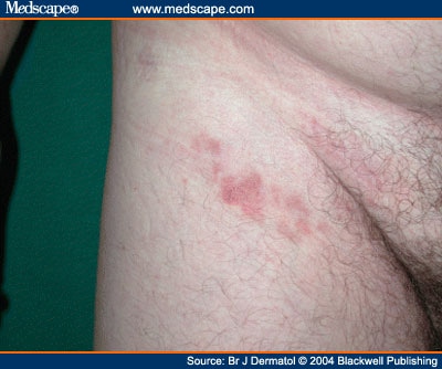
Early Patch Stage Mycosis Fungoides

Cutaneous T-cell lymphoma, also known as mycosis fungoides, is a malignancy of the T helper. Since the early patch stage may mimic chronic inflammatory dermatoses. FIGURE 1 Clinical manifestations of mycosis fungoides —Image (A) shows typical early patch with erythema and mild scale; (B) shows a typical plaque, with raised.
What is Mycosis Fungoides? The disease condition of mycosis fungoides is an example of a blood cancer called as cutaneous T-cell lymphoma. Bitdefender Internet Security 2008 V11.0.15 Keygen(patch). In this malignancy, the T-cells of the blood become cancerous and affects the skin. The result is the appearance of different types of skin lesion.
Although the lesions are on the skin, the skin cells do not become malignant [3, 4]. Figure 1 shows an example of lesions associated with mycosis fungoides. Figure 1- Mycosis fungoides The prognosis of this condition depends on several factors such as the age of the patient and the progression of the condition. Those who were diagnosed at the early phase of mycosis fungoides have a 5-year survival rate of 100% while those in the last phase of the condition have a 5-year survival rate of 14% [5]. Phases The condition have a slow progression and undergoes several distinct stages. It is important to note that not everyone will undergo through all of the stages [1, 2, 6]. Premycotic phase Most patients will develop flat and scaly lesions that are reddish or pinkish.
Form 433 D Installment Agreement. The malignant T-cells are present in these lesions. These do not cause any symptoms and may last for several months or even years [1, 2, 6]. Patch phase Eczema-like patches will appear in the body. They are commonly found in upper thighs, lower abdomen, breasts and buttocks [1, 2, 6].
Plaque phase The plaques are reddish or brownish raised lesions. These plaques may develop from the patches or they may develop on their own. There are patients who have patches and plaques at the same time [1, 2, 6]. Tumor phase Tumors that are deeper and thicker than plaques may develop. As before, the tumors may develop from existing patches and plaques or they can form on their own. There are times that these tumors will have open sores which increases the risk for infection.
The cancerous T-cells may spread to other organs such as the lungs, liver, spleen and lymph nodes [1, 2, 6].
Plaque of mycosis fungoides Typical visible symptoms include rash-like patches, tumors,. Itching () is common, perhaps in 20 percent of patients, but is not universal.
The symptoms displayed are progressive, with early stages consisting of lesions presented as scaly patches. These lesions prefer the buttock region. The later stages involve the patches evolving into plaques distributed over the entire body. The advanced stage of mycosis fungoides is characterized by generalized, with severe pruritus and scaling. Cause [ ] The cause of mycosis fungoides is unknown, but it is not believed to be hereditary or genetic in the vast majority of cases. One incident has been reported of a possible genetic link.